Home

טוב כדורגל להפריע gfp laser זברה דרום אמריקה מאולץ

Green Fluorescent Protein - Cool Uses - Living Laser

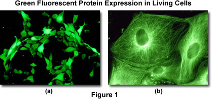
Confocal Microscopy - Green Fluorescent Protein | Olympus LS

Ultralow-threshold green fluorescent protein laser based on high Q  microbubble resonators
Ultralow-threshold green fluorescent protein laser based on high Q microbubble resonators

5 Ways to Improve Your Fluorescent Protein Sorting

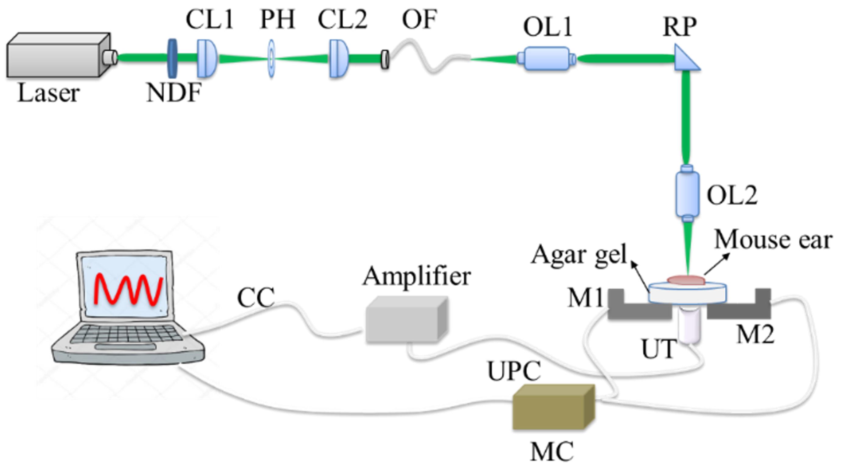
Sensors | Free Full-Text | Dual-Modal In Vivo Fluorescence/Photoacoustic  Microscopy Imaging of Inflammation Induced by GFP-Expressing Bacteria
Sensors | Free Full-Text | Dual-Modal In Vivo Fluorescence/Photoacoustic Microscopy Imaging of Inflammation Induced by GFP-Expressing Bacteria

Fluorescent Proteins- Flow Cytometry Core Facility

Single GFP-expressing cell is basis of a living biological laser device |  RobAid
Single GFP-expressing cell is basis of a living biological laser device | RobAid

Feasibility, sensitivity, and reliability of laser-induced fluorescence  imaging of green fluorescent protein-expressing tumors in vivo: Molecular  Therapy
Feasibility, sensitivity, and reliability of laser-induced fluorescence imaging of green fluorescent protein-expressing tumors in vivo: Molecular Therapy

GFP fluorescence peak fraction analysis based nanothermometer for the  assessment of exothermal mitochondria activity in live cells | Scientific  Reports
GFP fluorescence peak fraction analysis based nanothermometer for the assessment of exothermal mitochondria activity in live cells | Scientific Reports

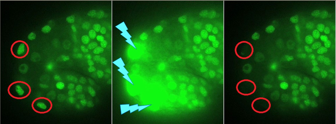
Application Of Laser Micro-Irradiation For Examination Of Single And Double  Strand Break Repair In Mammalian Cells - Video
Application Of Laser Micro-Irradiation For Examination Of Single And Double Strand Break Repair In Mammalian Cells - Video
Team uses lasers to spatially and temporally control the release of a  tagged protein inside a cell
Team uses lasers to spatially and temporally control the release of a tagged protein inside a cell

GFP helps turn single cell into “living laser” | BioScan | Sep 2011 |  BioPhotonics
GFP helps turn single cell into “living laser” | BioScan | Sep 2011 | BioPhotonics

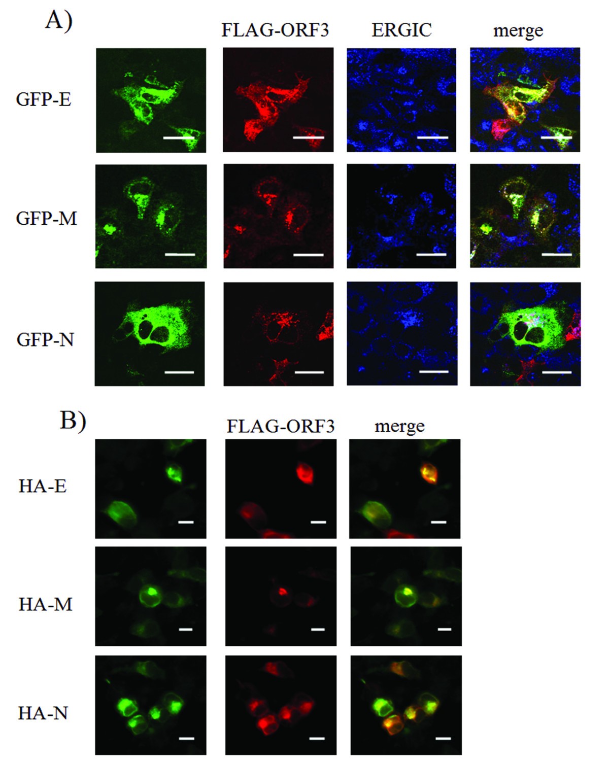
Human Coronavirus NL63 Open Reading Frame 3 encodes a virion-incorporated  N-glycosylated membrane protein | Virology Journal | Full Text
Human Coronavirus NL63 Open Reading Frame 3 encodes a virion-incorporated N-glycosylated membrane protein | Virology Journal | Full Text
A Photoactivatable GFP for Selective Photolabeling of Proteins and Cells |  Science
A Photoactivatable GFP for Selective Photolabeling of Proteins and Cells | Science

Fluorescent Proteins and the Story Behind GFP

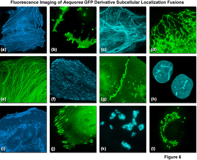
ZEISS Microscopy Online Campus | Jellyfish Fluorescent Proteins

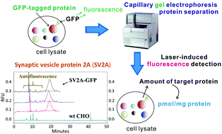
Quantification of green fluorescent protein-(GFP-) tagged membrane proteins  by capillary gel electrophoresis - Analyst (RSC Publishing)
Quantification of green fluorescent protein-(GFP-) tagged membrane proteins by capillary gel electrophoresis - Analyst (RSC Publishing)

Quantitative and time-resolved monitoring of organelle and protein delivery  to the lysosome with a tandem fluorescent Halo-GFP reporter | Molecular  Biology of the Cell
Quantitative and time-resolved monitoring of organelle and protein delivery to the lysosome with a tandem fluorescent Halo-GFP reporter | Molecular Biology of the Cell

Single GFP-expressing cell is basis of living | EurekAlert!

Green fluorescent protein - Wikipedia

DsRed as a Potential FRET Partner with CFP and GFP: Biophysical Journal

Laser Information for Mosaic Photostimulation- Oxford Instruments

Fig. 1.8, Transfected cells expressed GFP and formed dome-shaped colonies  on days 4–6 following laser transfection - Optically Induced Nanostructures  - NCBI Bookshelf
Fig. 1.8, Transfected cells expressed GFP and formed dome-shaped colonies on days 4–6 following laser transfection - Optically Induced Nanostructures - NCBI Bookshelf

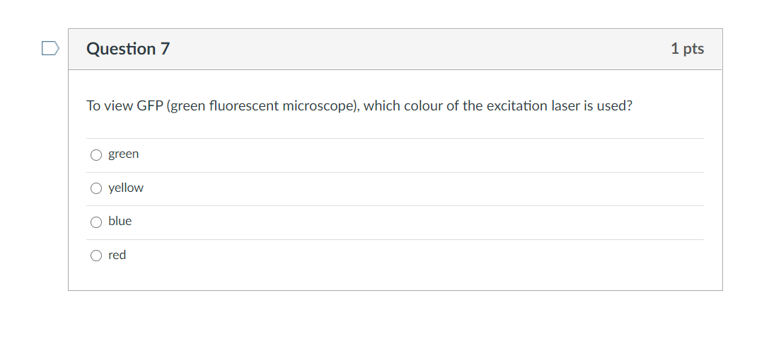
Solved Question 7 1 pts To view GFP (green fluorescent | Chegg.com

Laser Capture Microdissection of Single Neurons with Morphological  Visualization Using Fluorescent Proteins Fused to Transmembrane Proteins |  eNeuro
Laser Capture Microdissection of Single Neurons with Morphological Visualization Using Fluorescent Proteins Fused to Transmembrane Proteins | eNeuro

Fluorescent Proteins- Flow Cytometry Core Facility